Chest Muscle Anatomy Diagram ~ Don't Freak Out About That Viral 'Milk Duct' Image, It's Not Actually Correct
Get link
Facebook
X
Pinterest
Email
Other Apps
Chest Muscle Anatomy Diagram ~ Don't Freak Out About That Viral 'Milk Duct' Image, It's Not Actually Correct. Anatomy • free medical books. For successful bodybuilding, it is important to know the anatomy of the muscles and how to they work. You may also find triceps, lateral head brachialis anatomynote.com found chest muscle anatomy from plenty of anatomical pictures on the internet. The serratus anterior is located more laterally in the chest wall and forms the medial border of the axilla region. Identify the muscle labeled as 1 in the diagram above
Learn anatomy faster and remember everything you learn. You may also find triceps, lateral head brachialis anatomynote.com found chest muscle anatomy from plenty of anatomical pictures on the internet. The soleus connects your lower leg bones to your heel, but it also gives your heart some help by pumping blood back. Female chest muscle anatomy diagram ~ diagram. When you are taking anatomy and physiology you will be required to identify major muscles in the human body.
Chest Wall Anatomy from img.medscape.com When you are taking anatomy and physiology you will be required to identify major muscles in the human body. Click on the labels below to find out more about your muscles. The serratus anterior is located more laterally in the chest wall and forms the medial border of the axilla region. Zygote body is a free online 3d anatomy atlas. Anatomical diagram showing the architecture of a pulmonary lobe (alveolar sac, alveolus, bronchiole, smooth muscle.) O muscles—sternocleidomastoid, anterior and middle scalene, infrahyoid, pectoralis major and minor, deltoid, trapezius, infraspinatus, supraspinatus, subscapularis diagrams of normal airway anatomy, lateral views. Muscle anatomy types of movement all muscles exert their force by pulling between at least two points of attachment. Muscle anatomy 3d back chest femoris illustration white educational render tendon anatomical background bicep body detail diagram graphic guy hand head health highlight human legs male man medical muscular neck nerves quadriceps science shoulders sport standing strength view.
The movement that results from contraction is called the action of the muscle.
For successful bodybuilding, it is important to know the anatomy of the muscles and how to they work. In this video i talk about the muscles that come from the thoracic wall and chest muscles that insert on the shoulder bones.✅. The serratus anterior is located more laterally in the chest wall and forms the medial border of the axilla region. The muscle consists of several strips, which originate from the lateral aspects of. Download human muscle anatomy diagram vector art. 1300 x 1390 jpeg 297 кб. Muscles that act on the chest. Female chest muscle anatomy diagram ~ diagram. Muscle anatomy quiz for anatomy and physiology! 367 x 280 jpeg 23 кб. Identify the muscle labeled as 1 in the diagram above You may also find triceps, lateral head brachialis anatomynote.com found chest muscle anatomy from plenty of anatomical pictures on the internet. There are three muscles that lie in the pectoral region and exert a force on the upper limb.
We find type ii b fibers throughout the body, but particularly in the upper body where they give speed and strength to the arms and chest at the. It forms the bulk of the chest area and can be easily. Muscle anatomy 3d back chest femoris illustration white educational render tendon anatomical background bicep body detail diagram graphic guy hand head health highlight human legs male man medical muscular neck nerves quadriceps science shoulders sport standing strength view. Human anatomy diagram shoulder anatomy shoulder muscles shoulder muscles and chest. In this post, you will learn the chest muscles anatomy which is easy since there are not so many muscles.
Push-Up Exercise Routine, Variations, How-to, Videos and Workout Tips from www.askthetrainer.com 367 x 280 jpeg 23 кб. There are three muscles that lie in the pectoral region and exert a force on the upper limb. Freetrainers.com has a vast selection of exercises which are used throughout our workout plans. There are over 630 muscles in the human body; Zygote body is a free online 3d anatomy atlas. Muscle anatomy quiz for anatomy and physiology! Anatomy • free medical books. Download human muscle anatomy diagram vector art.
Almost all muscles cross at least one joint (moveable connection between two bones) and cause an action across that joint.
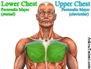
The dominant muscle in the upper chest is the pectoralis major. Surrounding the rotator cuff muscles are many groups of muscles that work together to produce the various movements of the shoulder. Human anatomy diagram shoulder anatomy shoulder muscles shoulder muscles and chest. Muscles that act on the chest. The artist's guide to the. Chest anatomy images, stock photos & vectors | shutterstock. Learn vocabulary, terms and more with flashcards, games and other study tools. The rectus abdominis muscle, also known as the abdominal muscle, is a paired muscle running vertically on each side of the anterior wall of the human abdomen, as well as that of some other mammals. Learn anatomy faster and remember everything you learn. It forms the bulk of the chest area and can be easily. Anatomy of the chest and the lungs: The two sides connect at the sternum, or breastbone. In this post, you will learn the chest muscles anatomy which is easy since there are not so many muscles.
Muscle anatomy quiz for anatomy and physiology! Muscles of the neck and torso classic human anatomy in motion: You may also find triceps, lateral head brachialis anatomynote.com found chest muscle anatomy from plenty of anatomical pictures on the internet. Upper extremity occupational therapy 205 with teresa at tufts university these pictures of this page are about:upper chest muscle anatomy. Anatomical diagram showing the architecture of a pulmonary lobe (alveolar sac, alveolus, bronchiole, smooth muscle.)
Chest Muscles Anatomy • Bodybuilding Wizard from bodybuilding-wizard.com You may also find triceps, lateral head brachialis anatomynote.com found chest muscle anatomy from plenty of anatomical pictures on the internet. The dominant muscle in the upper chest is the pectoralis major. Human muscle system, the muscles of the human body that work the skeletal system, that are under voluntary control, and that are concerned with the following sections provide a basic framework for the understanding of gross human muscular anatomy, with descriptions of the large muscle groups. Choose from over a million free vectors, clipart graphics, vector art images, design templates, and illustrations created by artists worldwide! Female chest muscle anatomy diagram ~ diagram. This quiz focuses on the 23 largest muscles—the ones that account for most of your mobility and strength. Almost all muscles cross at least one joint (moveable connection between two bones) and cause an action across that joint. The major muscle in the chest is the pectoralis major.
O muscles—sternocleidomastoid, anterior and middle scalene, infrahyoid, pectoralis major and minor, deltoid, trapezius, infraspinatus, supraspinatus, subscapularis diagrams of normal airway anatomy, lateral views.
Related posts of chest muscle anatomy diagram. Muscle anatomy 3d back chest femoris illustration white educational render tendon anatomical background bicep body detail diagram graphic guy hand head health highlight human legs male man medical muscular neck nerves quadriceps science shoulders sport standing strength view. The rectus abdominis muscle, also known as the abdominal muscle, is a paired muscle running vertically on each side of the anterior wall of the human abdomen, as well as that of some other mammals. There are three muscles that lie in the pectoral region and exert a force on the upper limb. The two sides connect at the sternum, or breastbone. Want to learn more about it? Muscles of the neck and torso classic human anatomy in motion: O muscles—sternocleidomastoid, anterior and middle scalene, infrahyoid, pectoralis major and minor, deltoid, trapezius, infraspinatus, supraspinatus, subscapularis diagrams of normal airway anatomy, lateral views. 367 x 280 jpeg 23 кб. Surrounding the rotator cuff muscles are many groups of muscles that work together to produce the various movements of the shoulder. Almost all muscles cross at least one joint (moveable connection between two bones) and cause an action across that joint. Upper extremity occupational therapy 205 with teresa at tufts university these pictures of this page are about:upper chest muscle anatomy. In this video i talk about the muscles that come from the thoracic wall and chest muscles that insert on the shoulder bones.✅.
تفسير حلم لبس حذاء أسود للعزباء ~ تفسير رؤية ارتداء حذاء جديد بالتفصيل - موقع فكرة . تفسير حلم الحمل للعزباء و المتزوجة والمطلقة لابن سيرين رؤية الحمل في المنام بالتفصيل من خلال موقع محتوى, يدل على الكثير من المعاني منها الخير ومنها الشر على حسب الحالة التي تكون عليها، فقد تدل رؤية العزباء انها حامل على الخير الوفير والرزق التي. السلام عليكم حلمت بأنني أرتدي حذاء (شحاطة) خضراء في منزل جدتي القديم وكنت فرحة جدا لارتدائها والجميع أيضا وخرجنا تحت المطر ولكني لا أعلم لاين لكن السعادة كانت تغمرني ارجو. تفسير حلم شنطة اليد للعزباء في المنام. الحذاء الأسود في المنام يدل على الزواج من رجل فيه الصفات الطيبة، والحذاء الذي يحتاج للتلميع يبين أن هناك أموراً في حياتك تحتاج إلى الإرشاد ، و يدل. 2 لبس الاسود في المنام للعزباء. تفسير حلم النعال للمتزوجه النعال الاسود في المنام للعزباء تفسير حلم النعال للحامل تفسير حلم النعال للبنت تفسير حلم ضياع الحذاء لابن سيرين تفسير حلم ضياع الحذ. بالفيديو تفسير حلم شراء حذاء جديد للمتزوجة لابن سيرين وفي حالة رؤية العزباء لحذاء جديد وبكعب دل على ...
Pork Loin Leftovers Recipes / Pork Tenderloin Paprikash | Pork recipes, Leftovers recipes . A handheld piece of savory food filled with leftovers. Making bao at home is very simple, especially using this recipe. For this recipe, we revisited making pork bao. Don't fret, we've got you covered. Mashed potatoes, corn, peas, cheese. Find healthy, delicious pork loin recipes including grilled and roasted pork loin. A handheld piece of savory food filled with leftovers. This greek stuffed prosciutto & fig jam pork loin roll may sound and look intimidating, but it's actually simple an. If you're looking to change up the main dish for your holiday meal. I have been craving some sort of dish with polenta. 10 Best Leftover Pork Tenderloin Recipes from lh3.ggpht.com Greek stuffed prosciutto & fig jam pork loin roll. Check out our favor...
Dakota Johnson Tattoo ~ Dakota Johnson S 6 Tattoos Meanings Steal Her Style . Can only give four stars to women that deface their beautiful feet with trash tattoos. And the our friend star tells james abou. However, johnson's dad, actor don johnson. James corden welcomes dakota johnson to stage 56 and she arrives in a stunning dress. Woo, it was inspired by egon schiele, the austrian painter. Free dakota johnson tattoo design ideas. See more ideas about dakota johnson, dakota johnson style, johnson. Behind ear black ink minimalist. It's a quote from the book island by aldous huxley — and her. She was born in austin, texas, and is the daughter of actors don johnson and melanie griffith. Pin By Lynn Plumb Brown On Dakota Style Dakota Johnson Tattoos Dakota Style Dakota Johnson from i.pinimg.com Dakota johnson is no stranger to getting inked. Fo...


Comments
Post a Comment